
Rib Cage Muscles Anatomy - Anyone Else Felt Like They Pulled One Of Their Intercostal Muscles During A Kettlebell Workout More Details In Comments Kettlebell : Comments lesson notes downloads 1.. In this video, we explore:1) the anatomy of the sternum2) the anatomy and differences between the three classes of ribs3) the anatomy and differences between. This muscle lies between the back of your rib cage and your shoulder blade near the subscapularis (one of the four rotator cuff muscles). The costal angle also marks the attachment for some of the deep back muscles to. It provides a strong framework onto which the muscles of the shoulder girdle, chest, upper abdomen and back can attach. The pain may occur immediately upon injury or develop slowly over time.
Human body rib cage anatomy anterior stock illustration 553801780 these pictures of this page are about:anatomical rib cage. Comments lesson notes downloads 1. The space between each rib is called the intercostal space, and there are 11 intercostal spaces in the thoracic cage, which are filled with nerves, lymph nodes, arteries, veins, and muscles.in fact, when you eat ribs at a restaurant, you're eating the intercostal muscles of an animal. The muscles below my rib cage keep hurting. This muscle lies between the back of your rib cage and your shoulder blade near the subscapularis (one of the four rotator cuff muscles).

It expands the lower rib cage and is considered to be the main inspiratory muscle.
The rib cage is a bony structure found in the chest (thoracic cavity). It forms the bony framework for breathing. This muscle lies between the back of your rib cage and your shoulder blade near the subscapularis (one of the four rotator cuff muscles). Lesson by stan prokopenko in anatomy of the human body. The costal angle also marks the attachment for some of the deep back muscles to. Rib cage, in vertebrate anatomy, basketlike skeletal structure that forms the chest, or thorax, and is made up of the ribs and their corresponding attachments to the sternum (breastbone) and the vertebral column.the rib cage surrounds the lungs and the heart, serving as an important means of bony protection for these vital organs.in total, the rib cage consists of the 12 thoracic vertebrae and. The ribs are attached to the breastbone, which is the. Rib 2 is thinner and longer than rib 1, and has two articular facets on the head as normal. Originate at the lower border of the rib, inserting into the superior border of the rib below. The back end is wide and open. In this image, you will find thoracic vertebrum, costochondral joint, costal cartilage, costal margin, costal arch, thoracic vertebrum, xiphoid process, xiphisternal joint, body, manubrial sternal joint, manubrium, the sternal notch in it. In spite of its resistance, the cage is dynamic, allowing pulmonary ventilation to. In most undergraduate anatomy courses, you will need to understand the key landmarks on a typical rib bone, as well as general information about the ribs.
The thoracic cage (rib cage) is the skeletal framework of the thoracic wall, which encloses the thoracic cavity. Register a new.com for just $9.99 for the first year and get everything you need to make your mark online — website builder, hosting, email, and more. 16 photos of the rib cage diagram with organs. Diagram of human body, liver rib cage, rib cage diagram labeled, rib cage diagram numbered, rib cage diaphragm, rib cage heart, rib cage organs anatomy, rib cage pain, stomach, diagram of human body, liver rib cage, rib cage diagram labeled, rib cage diagram numbered, rib cage diaphragm, rib cage. Elevates the ribs, increasing the thoracic volume.

The rib cage is a bony structure found in the chest (thoracic cavity).
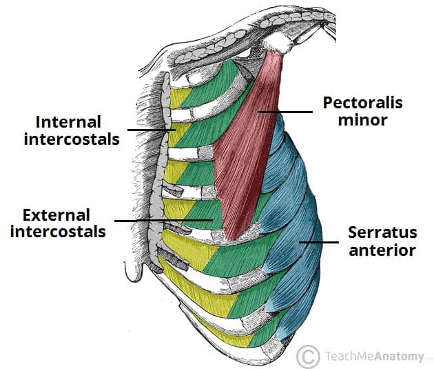
There are 11 pairs of external intercostal muscles. The upper edge is round and the lower sharp. In this rib bones anatomy quiz, you can test your knowledge of the ribs. In spite of its resistance, the cage is dynamic, allowing pulmonary ventilation to. Each pair is numbered based on their attachment to the sternum, a bony process at the front of the rib cage which serves as an anchor point. The rectus abdominis runs between the ribs and the pubic bone and supports movements between the rib cage and the pelvis. They run inferoanteriorly from the rib above to the rib below, and are continuous with the external oblique of the abdomen. Costae) are long, flat, curved bones that form the rib cage.there are twelve pairs of ribs, all of which articulate with the vertebral column, while only the first seven ribs directly articulate with the sternum.the rib cage forms the majority of the thoracic skeleton and provides protection for the internal thoracic organs, including the lungs and the heart. It is made up of 12 pairs of ribs. The rib cage has three important functions: Originate at the lower border of the rib, inserting into the superior border of the rib below. Elevates the ribs, increasing the thoracic volume. With the upper ribs, closer to the nodule (and in the case of lower ribs, a little further from the nodule) they are curved and have a rough surface that connects them with muscles, angulus costae.
Shaped somewhat like a cone, it is created by the individual ribs connecting to the spine above and to the sternum below. The ribs are attached to the breastbone, which is the. The body, or shaft, of the rib is thin, flat and curved. The pain may occur immediately upon injury or develop slowly over time. The major muscles of the body, anterior view.
In this rib bones anatomy quiz, you can test your knowledge of the ribs.
The right scapula from the front and back side. Quizzes are the secret to your success! It has a roughened area on its upper surface, from which the serratus anterior muscle originates. Costae) are long, flat, curved bones that form the rib cage.there are twelve pairs of ribs, all of which articulate with the vertebral column, while only the first seven ribs directly articulate with the sternum.the rib cage forms the majority of the thoracic skeleton and provides protection for the internal thoracic organs, including the lungs and the heart. The curve becomes most prominent at the costal angle, which is when the rib turns anterolaterally. The dome shaped thoracic cage provides the necessary rigidity for organ protection, weight support for the upper limbs and anchorage for muscles. Diagram of human body, liver rib cage, rib cage diagram labeled, rib cage diagram numbered, rib cage diaphragm, rib cage heart, rib cage organs anatomy, rib cage pain, stomach, diagram of human body, liver rib cage, rib cage diagram labeled, rib cage diagram numbered, rib cage diaphragm, rib cage. In spite of its resistance, the cage is dynamic, allowing pulmonary ventilation to. In this rib bones anatomy quiz, you can test your knowledge of the ribs. This muscle lies between the back of your rib cage and your shoulder blade near the subscapularis (one of the four rotator cuff muscles). The thoracic cage (rib cage) is the skeletal framework of the thoracic wall, which encloses the thoracic cavity. It is innervated by the first four lumbar nerves, plus the twelfth thoracic nerve. Human body rib cage anatomy anterior stock illustration 553801780 these pictures of this page are about:anatomical rib cage.
Register a newcom for just $999 for the first year and get everything you need to make your mark online — website builder, hosting, email, and more rib cage muscles. The major muscles of the body, anterior view.